


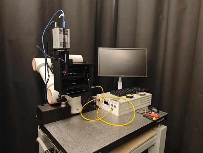
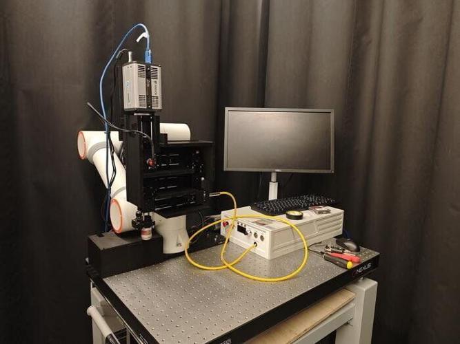


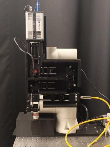


The embedded SMEAR-ULM system on a robotic arm and a movable utility cart for autonomous and precise positioning in Jinyang Liang's laboratory at the INRS. (Jinyang Liang via SWNS)
By Stephen Beech
A new "intelligent tattoo" is able to detect the deadliest form of skin cancer before it appears.
The "minimally invasive" technology has been shown to work in preclinical studies and could transform the early detection of tiny malignant tumors, say scientists.
Detecting melanoma before it becomes visible is a major challenge for dermatologists.
But researchers in Canada have developed a promising solution, already successfully tested on mice.
The high-tech system, called SMEAR-ULM, detects skin cancers at their earliest stages by measuring tiny temperature variations at the surface of the skin.
The study, published in the journal Nature Sensors, was conducted by scientists from the University of Montreal and the University of Quebec's National Institute for Scientific Research (INRS).
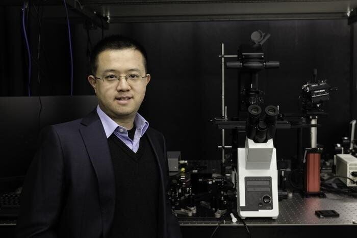
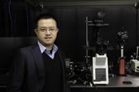
Professor Jinyang Liang, specializing in ultrafast imaging and biophotonics at INRS, has developed with colleagues at INRS and Université de Montréal a minimally invasive technology to detect skin cancer before it appears. Called SMEAR-ULM, it’s a high-tech system that can detect skin cancers at their earliest stages by measuring tiny temperature variations at the surface of the skin. (INRS via SWNS)
Research leader Jinyang Liang, of the INRS, says the work could have a "significant" impact on the detection of skin cancer.
Melanoma cases are on the rise with latest figures showing that, in 2022, a record number of more than 20,000 people in the UK were diagnosed with melanoma, the most serious form of skin cancer.
The figure is expected to rise to over 26,000 a year by 2040, according to Cancer Research UK.
Study senior author Liang says early diagnosis is "critical" to improving survival rates.
But current diagnostic approaches rely on visual examination followed by biopsies — procedures that are invasive and sometimes unnecessary.
By enabling rapid, direct, and non-invasive assessment of suspicious skin lesions, Liang says the new technology could reduce unnecessary biopsies, improve early diagnostic accuracy, and support clinical decision-making.

Yingming Lai, an INRS postdoctoral fellow who completed his PhD in Energy and Materials Sciences at INRS in Jinyang Liang's laboratory. (INRS via SWNS)
He said: "Our goal is to provide a minimally invasive tool to detect very small, but still aggressive melanomas.
"Because of their small size, the melanomas are usually excluded from clinical visual inspection, which leaves the threat unwatched.
"We want to detect them, so that intervention can be made as soon as possible."
Study co-corresponding author Sylvain Meloche, from the University of Montreal, said: "Even though this study was conducted in mice, this animal model replicates the genetic changes observed in human melanomas and could therefore potentially benefit patients."
The researchers explained that the approach also redefines the role of temperature in cancer detection.
While tumors are known to generate more heat due to their higher metabolic activity, the signal has traditionally been too imprecise to use as a diagnostic marker.

The 10-inch × 10-inch × 2-inch module of the SMEAR-ULM system. (Jinyang Liang via SWNS)
However, SMEAR-ULM changes that by turning subtle thermal variations into a highly sensitive and measurable signal.
Liang, who specializes in ultrafast imaging and biophotonics at INRS, said: "At the core of the system is a patch of painless microneedles that deposits specialized nanoparticles just beneath the skin.
"These nanoparticles form a temporary 'intelligent tattoo' that behaves like an array of microscopic thermometers.
"When illuminated with near-infrared light, the nanoparticles emit visible light.
"Crucially, the lifetime of this light emission — how long it lasts — depends directly on local temperature.
"Because cancer cells consume more oxygen and nutrients than healthy cells, they produce additional heat, which can be detected through this optical signal."
Using an ultrafast imaging system, SMEAR-ULM captures all that information in a single high-speed snapshot, generating a detailed thermal map with sub-millimeter spatial resolution and sub-degree temperature sensitivity.
Study first author Yingming Lai, of INRS, said: "We capture all the necessary information for an instantaneous temperature map in a single shot, which makes the method fast and robust to continuously monitor abnormal thermal responses in small melanomas — even within complex in vivo conditions."
Using the new approach, the research team successfully detected micro-melanomas as early as four days old — a stage at which they are usually far too small to be identified by conventional imaging techniques.

Sylvain Meloche, a researcher at UdeM's Institute for Research in Immunology and Cancer and co-corresponding author of the study published in Nature Sensors. (Jinyang Liang via SWNS)
Conversely, conventional thermal imaging methods rely on infrared technologies that suffer from limited spatial resolution and high noise levels.
As a result, they usually only detect tumors larger than five millimeters (0.2 inches) — lesions already visible to the naked eye.
Current microneedle-based sensing approaches also require repeated measurements, limiting their use in living subjects.
The Canadian researchers say the SMEAR-ULM technology overcomes those limitations by combining microneedle encoding, rare-earth-doped nanoparticles, and ultrafast optical imaging into a system capable of real-time, single-shot thermal mapping in vivo.
Liang added: "This breakthrough effectively transforms skin temperature from a secondary indicator into a precise diagnostic biomarker for early-stage melanoma."
He believes that, beyond skin cancer detection, the platform could be adapted to map other physiological parameters — such as acidity concentrations — opening new possibilities in biomedical imaging and diagnostics.